Types Of Eyeball
The eyeball is a complex and fascinating organ responsible for our vision. The human eye is a spherical structure that is composed of various tissues, including the sclera, cornea, iris, pupil, lens, and retina. The eyeball is protected and supported by muscles, blood vessels, and other structures that help it function properly.
Understanding the different types of eyeball is important for gaining insights into how they work and how they can be affected by various conditions and disorders. The anatomical structure of the eyeball is a crucial aspect of its function, and it is made up of several components that work together to produce vision. The optical components of the eyeball, including the lens and retina, play a key role in the formation of images, while the protective and supporting structures help to maintain the integrity of the eye.
Key Takeaways
- The anatomical structure of the eyeball is composed of various tissues and structures that work together to produce vision.
- The optical components of the eye, including the lens and retina, play a crucial role in the formation of images.
- Protective and supporting structures help maintain the integrity of the eye, and understanding the different types of eyeball is important for gaining insights into how they function and how they can be affected by various conditions and disorders.
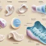
Anatomical Structure of the Eyeball
The eyeball is a complex organ that is responsible for vision. It is composed of three layers: the fibrous layer, the vascular layer, and the inner layer. Each layer has a specific function that contributes to the overall function of the eye.
Fibrous Layer
The fibrous layer is the outermost layer of the eyeball. It is composed of two parts: the sclera and the cornea. The sclera is the white, opaque part of the eye that covers most of the eyeball. It provides protection and support for the eyeball. The cornea is the clear, dome-shaped surface that covers the front of the eye. It is responsible for refracting light and focusing it onto the retina.
Vascular Layer
The vascular layer is also known as the uvea and is located beneath the fibrous layer. It is composed of three parts: the choroid, the ciliary body, and the iris. The choroid is a dark, pigmented layer that lines the inside of the sclera. It provides nourishment to the retina. The ciliary body is responsible for producing aqueous humor, which helps maintain the shape of the eye. The iris is the colored part of the eye that regulates the amount of light that enters the eye.
Inner Layer
The inner layer is the innermost layer of the eyeball. It is composed of two parts: the retina and the optic nerve. The retina is a thin layer of tissue that lines the back of the eye. It contains photoreceptor cells that convert light into electrical signals that are transmitted to the brain. The optic nerve is a bundle of nerve fibers that carries these signals from the retina to the brain.
Other important structures of the eyeball include the lens and the vitreous body. The lens is a transparent structure located behind the iris that helps focus light onto the retina. The vitreous body is a clear, gel-like substance that fills the space between the lens and the retina. It helps maintain the shape of the eyeball and provides support for the retina.
In summary, the anatomical structure of the eyeball is a complex and intricate system that allows for the perception of light and the formation of visual images. Each layer and structure has a specific function that contributes to the overall function of the eye.
Optical Components and Vision
Refraction and Focusing
The eyeball is a complex optical instrument that allows us to see the world around us. The cornea and lens are the primary optical components of the eye. The cornea is the clear, outermost layer of the eye that acts as a protective covering. The lens is a transparent, flexible structure that changes shape to focus light onto the retina.
When light enters the eye, it passes through the cornea and then through the pupil, which is the opening in the center of the iris. The iris is the colored part of the eye that controls the size of the pupil. The lens then changes shape to focus the light onto the retina at the back of the eye.
Photoreception and Image Processing
The retina is the innermost layer of the eye and contains millions of photoreceptor cells called rods and cones. Rods are responsible for detecting light and dark, while cones are responsible for color vision. The macula is a small area in the center of the retina that contains a high concentration of cones and is responsible for sharp, detailed vision. The fovea is a small depression in the center of the macula where visual acuity is highest.
When light strikes the photoreceptor cells in the retina, it triggers a series of chemical reactions that generate electrical signals. These signals are then transmitted to the brain via the optic nerve. The brain then processes these signals to create the visual perception of the world around us.
In summary, the eyeball is a complex optical instrument that allows us to see the world around us. The cornea and lens focus light onto the retina, where photoreceptor cells detect the light and transmit signals to the brain. The brain then processes these signals to create the visual perception of the world around us.
Protective and Supporting Structures
The eyeball is a delicate organ that is protected and supported by various structures. These structures include the orbital cavity, extraocular muscles, eyelids, and tear system.
Orbital Cavity
The orbital cavity is a bony socket that houses the eyeball and its associated structures. The orbital cavity is formed by several bones, including the frontal bone, maxilla, zygomatic bone, and sphenoid bone. The orbital cavity provides protection to the eyeball from external forces and also helps to maintain the position of the eyeball.
Extraocular Muscles
The extraocular muscles are a group of six muscles that attach to the eyeball and control its movement. These muscles include the superior rectus, inferior rectus, medial rectus, lateral rectus, superior oblique, and inferior oblique muscles. The extraocular muscles work together to move the eyeball in various directions and maintain its position.
Eyelids and Tear System
The eyelids and tear system are responsible for protecting and lubricating the eyeball. The eyelids consist of the upper and lower eyelids, which protect the eyeball from foreign objects and help to distribute the tear film across the surface of the eyeball. The tear system includes the lacrimal gland, which produces tears, and the tear ducts, which drain excess tears from the eye.
The tear film is a thin layer of fluid that covers the surface of the eyeball and helps to keep it moist and lubricated. The tear film is composed of three layers: the lipid layer, aqueous layer, and mucin layer. The lipid layer is produced by the meibomian glands in the eyelids and helps to prevent evaporation of the tear film. The aqueous layer is produced by the lacrimal gland and provides nutrients and oxygen to the cornea. The mucin layer is produced by the goblet cells in the conjunctiva and helps to spread the tear film evenly across the surface of the eyeball.
In conclusion, the protective and supporting structures of the eyeball play a crucial role in maintaining the health and function of the eye. These structures work together to protect the eyeball from external forces, control its movement, and keep it moist and lubricated.
Common Eye Conditions and Disorders
The human eye is a complex organ that can be affected by a variety of conditions and disorders. Some of the most common conditions are refractive errors, cataracts, glaucoma, and retinal disorders.
Refractive Errors
Refractive errors are a common type of eye problem that affect many people. These errors occur when the shape of the eyeball or the curvature of the cornea prevents light from being focused properly on the retina. This can lead to blurred vision, headaches, and eye strain. The most common types of refractive errors are myopia (nearsightedness), hyperopia (farsightedness), and astigmatism.
Cataracts and Glaucoma
Cataracts and glaucoma are two of the most common eye diseases in the world. Cataracts occur when the lens of the eye becomes cloudy, making it difficult to see clearly. Glaucoma is a condition that damages the optic nerve, which can lead to vision loss.
Retinal Disorders
Retinal disorders are a group of eye conditions that affect the retina, the layer of tissue at the back of the eye that is responsible for detecting light and sending visual signals to the brain. Some common retinal disorders include retinal detachment and macular degeneration.
It is important to note that many eye conditions and disorders can be treated with medications, surgery, or other interventions. Early detection and treatment can help to preserve vision and prevent further damage. Regular eye exams are an important part of maintaining good eye health and preventing serious eye problems.
Physiology of Vision
The proper function of the eye depends on its ability to receive and process energy from light in the environment, produce action potentials in specialized nerve cells, and relay those potentials through the optic nerve (cranial nerve II) to the brain. The physiology of vision can be divided into two main categories: light and color perception, and visual pathways and processing.
Light and Color Perception
Light is the primary stimulus for vision. The eye receives light through the pupil, which is controlled by the aperture, a ring of muscle that adjusts the size of the pupil to regulate the amount of light entering the eye. The pupillary light reflex is a reflex that controls the size of the pupil in response to changes in light intensity.
Color perception is the ability of the eye to distinguish between different wavelengths of light. The retina contains photoreceptor cells called rods and cones, which are responsible for detecting light and color. Rods are more sensitive to low levels of light and are responsible for peripheral vision, while cones are responsible for color vision and are concentrated in the center of the retina.
Visual Pathways and Processing
The visual pathway begins at the retina and continues through the optic nerve, optic chiasm, and optic tracts to the visual cortex in the brain. The optic chiasm is the point where the optic nerves from each eye cross over to the opposite side of the brain. This allows for binocular vision, which is the ability to perceive depth and three-dimensional space.
Visual processing occurs in several areas of the brain, including the primary visual cortex, which is responsible for basic visual processing, and higher visual areas, which are responsible for more complex visual processing, such as object recognition and facial recognition.
Depth perception is the ability to perceive the distance of objects in the environment. This is accomplished through several cues, including binocular disparity, which is the difference in the images seen by each eye, and monocular cues, which are visual cues that can be perceived with one eye.